- Online Store
- >
- Image Atlas Series
- >
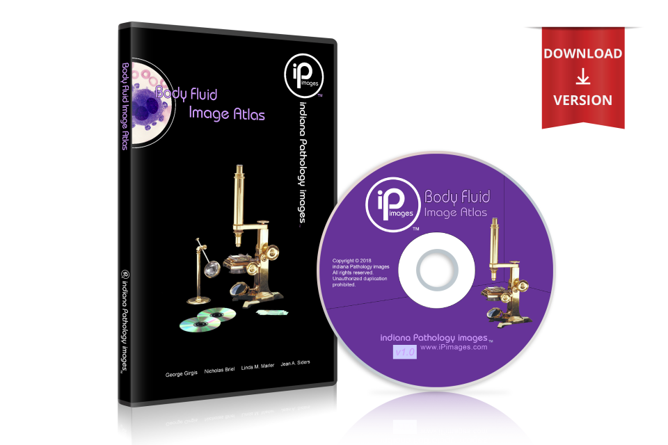
- Body Fluid Image Atlas - Single User (Download)
Body Fluid Image Atlas - Single User (Download)
SKU:
$175.00
$175.00
Unavailable
per item
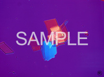
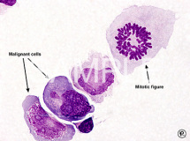
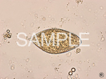
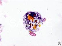
- over 700 high-quality images
- images easily downloaded into presentation programs (e.g. PowerPoint®)
- effortless printing for exams and manuals
- Easy-to-use web-based format
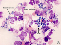
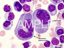
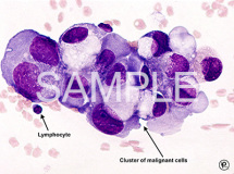
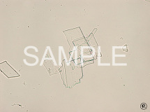
- Crystals, Granules, Lining cells, Malignant cells, Microorganisms, WBCs
- Fluids include: BALs, CSF, Cyst, Dialysate, Serous, Synovial, etc.
- Cell Index or Disease/Condition retrieval
- PC and Mac compatible
Image Atlas Series:
Each atlas contains a comprehensive collection of clinically relevant images in a click-art format. Images are high quality for viewing and printing purposes, and appropriately sized for easy use in most software programs. Each atlas of images will eliminate the need to scan pictures and slides or search the internet for teaching materials. Every title will be an invaluable educational resource and is certain to enhance any image library.
Suggested uses for Indiana Pathology Images™ products
Educators:
System requirements:
Windows 95, 98, NT, 2000, XP or 7-10
Mac OS X v.10.2 or above
Each atlas contains a comprehensive collection of clinically relevant images in a click-art format. Images are high quality for viewing and printing purposes, and appropriately sized for easy use in most software programs. Each atlas of images will eliminate the need to scan pictures and slides or search the internet for teaching materials. Every title will be an invaluable educational resource and is certain to enhance any image library.
Suggested uses for Indiana Pathology Images™ products
Educators:
- PowerPoint lectures
- Color images for examinations
- Student laboratory manuals
- Intranet web-site postings
- Intranet postings for review of lecture material
- At-home review
- Printed 'flash cards' for national certification examination review
- Source for reference material
- Review material for continuing education credit
- Training and retraining of employees
- Images that can be incorporated into laboratory procedure manuals
System requirements:
Windows 95, 98, NT, 2000, XP or 7-10
Mac OS X v.10.2 or above
Cell Index
Crystals
Calcium pyrophosphate dehydrate (CPPD)
Cholesterol
Hematoidin
Monosodium urate (MSU)
Steroid
Granules
Azurophilic granule
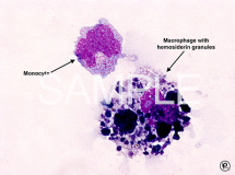
Hemosiderin granule
Lining Cells
Alveolar lining cell
Bronchial lining cell
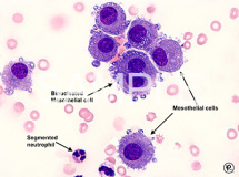
Mesothelial cell
Squamous epithelial cell
Synovial lining cell
Ventricular Lining (Ependymal/Choroid Plexus) cell
Malignant cells
Malignant cell
Mitotic figure
Microorganisms
Bacteria, bacilli
Bacteria, cocci
Fungus, Aspergillus
Fungus, Blastomyces
Fungus, Candida
Fungus, Cryptococcus
Fungus, Histoplasma
Fungus, Pneumocystis
Fungus, Yeast (not identified)
Parasite, Schistosoma hematobium
Parasite, Strongyloides
Parasite, Toxoplasma
Virus, Cytomegalovirus
Miscellaneous
Artifact
Capillary
Cartilage cell (Chondrocyte)
Corpora amylacea
Cytarabine within liposomes
Hemotoxylin body
Neural tissue
Red Blood Cells
Red blood cell
Red blood cell, nucleated
White Blood Cells
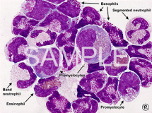
Basophil
Blast
Eosinophil
Erythrophage
LE (Lupus erythematosus) cell
Lymphocyte
Lymphocyte, reactive
Lymphoma cell
Macrophage
Macrophage with hematoidin crystal
Macrophage with hemosiderin granule (siderophage)
Macrophage with phagocytized red blood cell (erythrophage)
Macrophage, alveolar
Macrophage, signet ring cell
Metamyelocyte
Monocyte
Monocyte/Macrophage
Myelocyte
Neutrophil
Neutrophil, band
Neutrophil, segmented
Plasma cell
Plasma cell, abnormal
Promonocyte
Promyelocyte
Pyknotic cell
Siderophage
Signet ring cell
Cytospin Examination (low power)
Scanning - Alveolar lining cell
Scanning - Bacteria, bacilli
Scanning - Bacteria, cocci
Scanning - Basophil
Scanning - Blast
Scanning - Bronchial lining cell
Scanning - Calcium pyrophosphate dehydrate (CPPD) crystals
Scanning - Capillary
Scanning - Eosinophil
Scanning - Fungus, Aspergillus
Scanning - Fungus, Cryptococcus
Scanning - Fungus, Yeast (not identified)
Scanning - Hematoxylin body
Scanning - LE (Lupus Erythematosus) cell
Scanning - Lymphocyte
Scanning - Lymphoma cell
Scanning - Macrophage
Scanning - Macrophage, alveolar
Scanning - Malignant cell
Scanning - Mesothelial cell
Scanning - Mitotic figure
Scanning - Monocyte
Scanning - Monocytes/Macrophages
Scanning - Monosodium urate (MSU) crystal
Scanning - Neural tissue
Scanning - Neutrophil, band
Scanning - Neutrophil, segmented
Scanning - Parasite, Schistosoma hematobium
Scanning - Plasma cell
Scanning - Plasma cell, abnormal
Scanning - Promonocyte
Scanning - Promyelocyte
Scanning - Pyknotic cell
Scanning - Red blood cell
Scanning - Synovial lining cell
Scanning - Ventricular Lining (Ependymal/Choroid Plexus) cell
Disease/Condition by fluid type
Bronchoalveolar lavage
Leukemia, acute promyelocytic (APL)
Normal
Pneumonia - See pulmonary disease
Pulmonary disease, Aspergillosis
Pulmonary disease, Blastomycosis
Pulmonary disease, Candidiasis
Pulmonary disease, Cryptococcosis
Pulmonary disease, Cytomegalovirus (CMV)
Pulmonary disease, fungal
Pulmonary disease, Histoplasmosis
Pulmonary disease, Pneumocystis
Pulmonary disease, Streptococcus pneumoniae
Pulmonary disease, Strongyloidiasis
Pulmonary disease, Toxoplasmosis
Cerebrospinal fluid
Bone marrow contamination
Head trauma
Hemorrhage, subarachnoid
Infection, shunt
Leukemia, acute lymphoblastic (ALL)
Leukemia, acute monoblastic
Leukemia, acute myeloid (AML)
Leukemia, chronic myelogenous (CML)
Lymphoma
Meningitis, bacterial
Meningitis, fungal
Meningitis, viral
Neoplasm
Neoplasm, plasma cell
Neoplasm, treatment
Normal
Shunt malfunction
Traumatic tap
Cyst fluid
Inflammation
Dialysate fluid
Allergic reaction
Peritonitis
Serous fluid: Exudate
Aspergillosis
Infection
Neoplasm, plasma cell
Peritoneal fluid: Malignant effusion
Peritonitis
Pleural fluid: Lymphoma
Pleural fluid: Malignant effusion
Pleural fluid: Neoplasm, plasma cell
Systemic Lupus Erythematosus (SLE)
Serous fluid: Transudate
Congestive heart failure
Liver cirrhosis
Pleural fluid: Congestive heart failure
Suprapubic bladder aspirate
Schistosomiasis
Synovial fluid
Arthritis
Arthritis, gout
Arthritis, pseudogout
Arthritis, rheumatoid
Arthritis, septic
Normal
Systemic Lupus Erythematosus (SLE)
×
Crystals
Calcium pyrophosphate dehydrate (CPPD)
Cholesterol
Hematoidin
Monosodium urate (MSU)
Steroid
Granules
Azurophilic granule
Hemosiderin granule
Lining Cells
Alveolar lining cell
Bronchial lining cell
Mesothelial cell
Squamous epithelial cell
Synovial lining cell
Ventricular Lining (Ependymal/Choroid Plexus) cell
Malignant cells
Malignant cell
Mitotic figure
Microorganisms
Bacteria, bacilli
Bacteria, cocci
Fungus, Aspergillus
Fungus, Blastomyces
Fungus, Candida
Fungus, Cryptococcus
Fungus, Histoplasma
Fungus, Pneumocystis
Fungus, Yeast (not identified)
Parasite, Schistosoma hematobium
Parasite, Strongyloides
Parasite, Toxoplasma
Virus, Cytomegalovirus
Miscellaneous
Artifact
Capillary
Cartilage cell (Chondrocyte)
Corpora amylacea
Cytarabine within liposomes
Hemotoxylin body
Neural tissue
Red Blood Cells
Red blood cell
Red blood cell, nucleated
White Blood Cells
Basophil
Blast
Eosinophil
Erythrophage
LE (Lupus erythematosus) cell
Lymphocyte
Lymphocyte, reactive
Lymphoma cell
Macrophage
Macrophage with hematoidin crystal
Macrophage with hemosiderin granule (siderophage)
Macrophage with phagocytized red blood cell (erythrophage)
Macrophage, alveolar
Macrophage, signet ring cell
Metamyelocyte
Monocyte
Monocyte/Macrophage
Myelocyte
Neutrophil
Neutrophil, band
Neutrophil, segmented
Plasma cell
Plasma cell, abnormal
Promonocyte
Promyelocyte
Pyknotic cell
Siderophage
Signet ring cell
Cytospin Examination (low power)
Scanning - Alveolar lining cell
Scanning - Bacteria, bacilli
Scanning - Bacteria, cocci
Scanning - Basophil
Scanning - Blast
Scanning - Bronchial lining cell
Scanning - Calcium pyrophosphate dehydrate (CPPD) crystals
Scanning - Capillary
Scanning - Eosinophil
Scanning - Fungus, Aspergillus
Scanning - Fungus, Cryptococcus
Scanning - Fungus, Yeast (not identified)
Scanning - Hematoxylin body
Scanning - LE (Lupus Erythematosus) cell
Scanning - Lymphocyte
Scanning - Lymphoma cell
Scanning - Macrophage
Scanning - Macrophage, alveolar
Scanning - Malignant cell
Scanning - Mesothelial cell
Scanning - Mitotic figure
Scanning - Monocyte
Scanning - Monocytes/Macrophages
Scanning - Monosodium urate (MSU) crystal
Scanning - Neural tissue
Scanning - Neutrophil, band
Scanning - Neutrophil, segmented
Scanning - Parasite, Schistosoma hematobium
Scanning - Plasma cell
Scanning - Plasma cell, abnormal
Scanning - Promonocyte
Scanning - Promyelocyte
Scanning - Pyknotic cell
Scanning - Red blood cell
Scanning - Synovial lining cell
Scanning - Ventricular Lining (Ependymal/Choroid Plexus) cell
Disease/Condition by fluid type
Bronchoalveolar lavage
Leukemia, acute promyelocytic (APL)
Normal
Pneumonia - See pulmonary disease
Pulmonary disease, Aspergillosis
Pulmonary disease, Blastomycosis
Pulmonary disease, Candidiasis
Pulmonary disease, Cryptococcosis
Pulmonary disease, Cytomegalovirus (CMV)
Pulmonary disease, fungal
Pulmonary disease, Histoplasmosis
Pulmonary disease, Pneumocystis
Pulmonary disease, Streptococcus pneumoniae
Pulmonary disease, Strongyloidiasis
Pulmonary disease, Toxoplasmosis
Cerebrospinal fluid
Bone marrow contamination
Head trauma
Hemorrhage, subarachnoid
Infection, shunt
Leukemia, acute lymphoblastic (ALL)
Leukemia, acute monoblastic
Leukemia, acute myeloid (AML)
Leukemia, chronic myelogenous (CML)
Lymphoma
Meningitis, bacterial
Meningitis, fungal
Meningitis, viral
Neoplasm
Neoplasm, plasma cell
Neoplasm, treatment
Normal
Shunt malfunction
Traumatic tap
Cyst fluid
Inflammation
Dialysate fluid
Allergic reaction
Peritonitis
Serous fluid: Exudate
Aspergillosis
Infection
Neoplasm, plasma cell
Peritoneal fluid: Malignant effusion
Peritonitis
Pleural fluid: Lymphoma
Pleural fluid: Malignant effusion
Pleural fluid: Neoplasm, plasma cell
Systemic Lupus Erythematosus (SLE)
Serous fluid: Transudate
Congestive heart failure
Liver cirrhosis
Pleural fluid: Congestive heart failure
Suprapubic bladder aspirate
Schistosomiasis
Synovial fluid
Arthritis
Arthritis, gout
Arthritis, pseudogout
Arthritis, rheumatoid
Arthritis, septic
Normal
Systemic Lupus Erythematosus (SLE)
Home | Contact Us | Titles | Software | Posters | Bench-aids | FAQs | Support
Indiana Pathology Images © 2016

